

Scanning NV Microscope for Laboratory Material Analysis
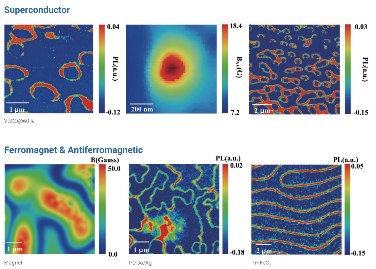
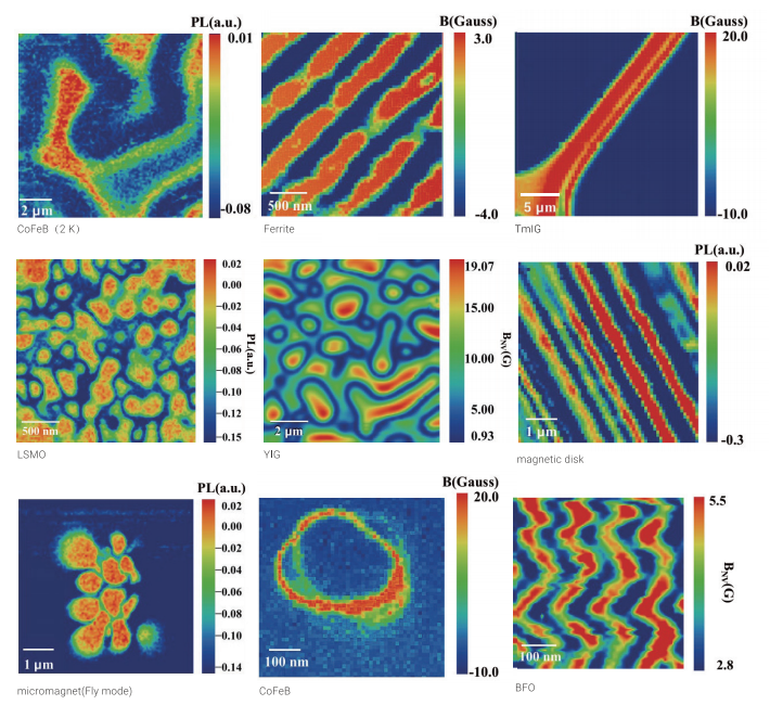
The Scanning NV Microscope is a quantum precision measurement instrument that combines optically detected magnetic resonance (ODMR) of diamond nitrogen-vacancy centers with scanning probe technique. It is applicable to research in spintronics, multiferroics, 2D magnetic materials, superconductors, and more.
Scanning NV Microscope Applications and Related Testing Solutions
Scanning NV Microscope supports precision inspection and analysis tasks that require clear imaging, stable test conditions, and dependable laboratory or production quality control.
Related Products: EASY-G 1310M Gas Pycnometer | Specific Surface & Porosimetry Analyzer EASY-V | Climber Series Specific Surface & Porosimetry Analyzer
evice Images:
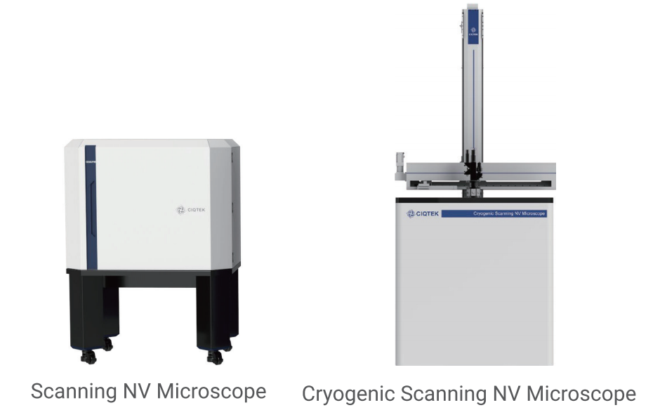
Left: Scanning NV Microscope (rectangular cabinet structure with four black feet).
Right: Cryogenic Scanning NV Microscope (taller white structure with slender top components).
Specifications Table:
Parameter | SNVM(RT) | SNVM(Cryogenic) |
|---|---|---|
Magnetic sensitivity | ≤2μT/√Hz/2 | ≤5μT/√Hz/2 |
Spatial resolution | 10–30 nm | 10–30 nm |
Temperature range | 300 K | 2 K–300 K |
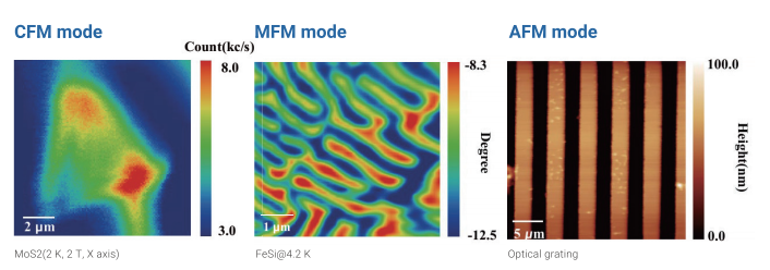
Main functions | ISO-B Mode, Dual-B Mode, Full-B Mode, Quenching Mode, AFM Mode, MFM Mode, CFM Mode | ISO-B Mode, Dual-B Mode, Full-B Mode, Quenching Mode, AFM Mode, MFM Mode, CFM Mode |
Magnet options | 0–50 mT | 9/1/1 T |
Key Components:
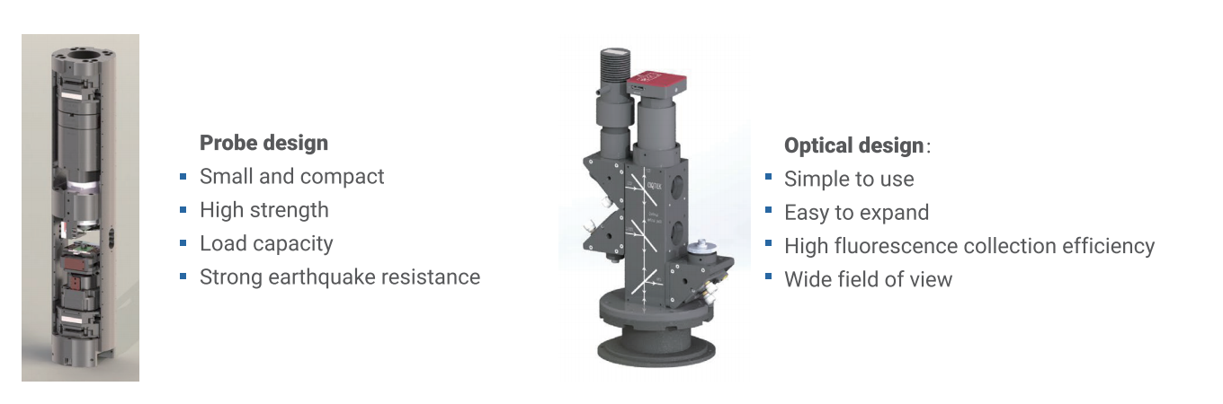
Probe Design: Small and compact strength; strong load capacity; strong earthquake resistance.
Optical Design: Simple to use; easy to expand; high fluorescence collection efficiency; wide field of view.
Applications:
Scanning NV probe with arrays of nanopillars or single pillar.
Count rates: ≥300 kc/s.
Contrast: ≥20%.
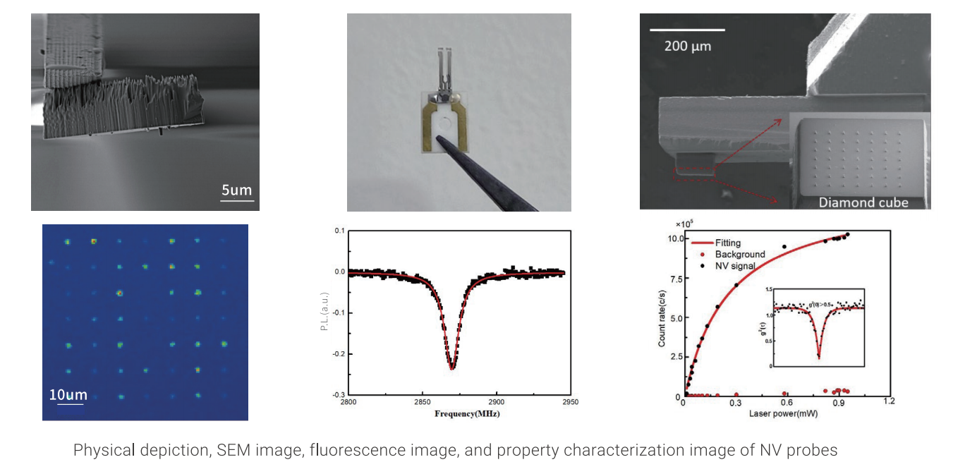
NV probe
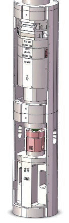
Device Diagram: Vertical cylindrical device for cryogenic high-pressure ODMR.
Technical Parameters Table: Parameter | Value |
|---|---|
Scan range | 300 μm × 300 μm |
Pressure | ≥30 GPa |
Magnetic sensitivity | ≤35μT/√Hz |
Spatial resolution | 500 nm |
Magnetic field | 9/1/1 T |
Refrigeration method | Cryogenic 1.5 W@4K |
Temperature range | 2 K–300 K |
Temperature resolution | ±50 mK |
Results Under 30 GPa:
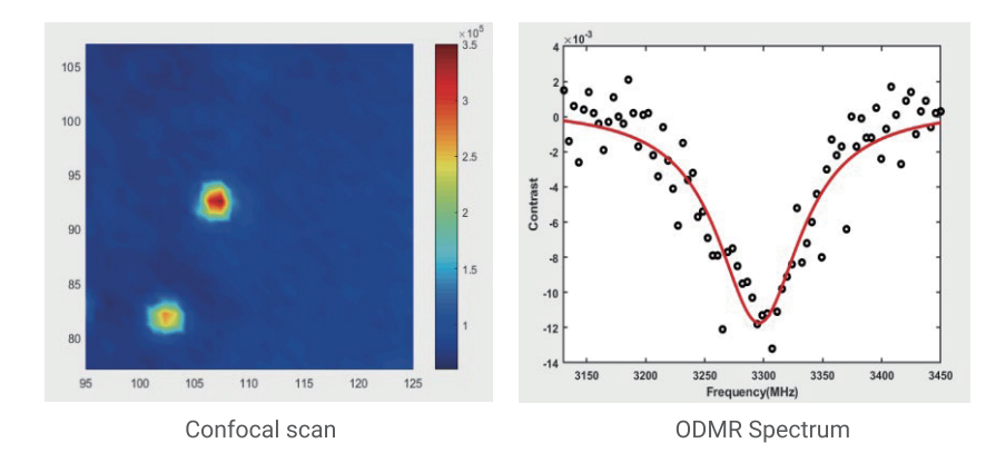
Confocal Scan: Blue background with two yellow-orange bright spots; color intensity scale included.
ODMR Spectrum: X-axis = frequency (MHz), Y-axis = contrast; black dots (experimental data), red curve (fitted model).
University List (Two Columns):
Left Column: Tsinghua University, City University of Hong Kong, Peking University, Institute of Physics CAS, University of Science and Technology of China, South China University ofudan University, Beihang University, Hebei University of Technology, Sun Yat-sen University, Harbin Institute of Technology, Xiamen University, Hubei Normal University.
Right Column: (Implied continuation of university names; exact list not fully specified but includes abbreviations).
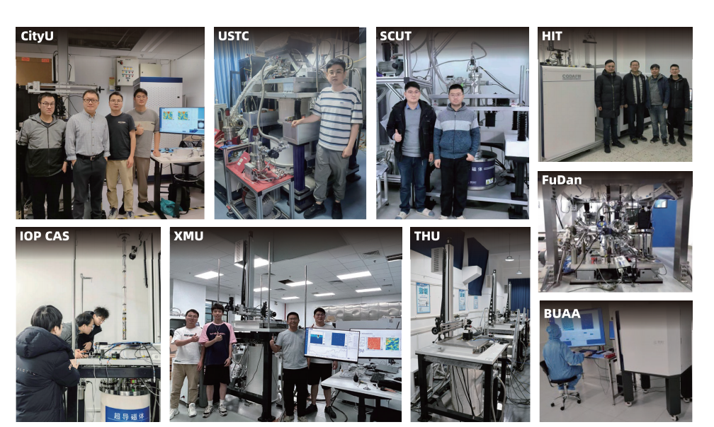
Research Scene Thumbnails (9 Images):
Abbreviations: CityU (City University of Hong Kong), USTC (University of Science and Technology of China), SCUT (South China University of Technology), HIT (Harbin Institute of Technology), FuDan (Fudan University), IOP CAS (Institute of Physics CAS), XMU (Xiamen University), THU (Tsinghua University), BUAA (Beihang University).
SEO Keywords
Scanning NV Microscope
Quantum Precision Measurement
Optically Detected Magnetic Resonance (ODMR)
Diamond Nitrogen-Vacancy Centers
Cryogenic Scanning NV Microscope
Magnetic Sensitivity
Spatial Resolution
NV Probe Superconductor
Cryogenic High Pressure ODMR
Confocal Scan
ODMR Spectrum
University Research Cases
Spintronics
Multiferroics
2D Magnetic Materials